
Welcome to the
Malaysian Family Physician, a peer-reviewed open-access journal of family practice
and primary care research.
TEST YOUR KNOWLEDGE
A CLERK WITH BACK PAIN
E Das Gupta FRCP; N Somaweera MD
International Medical University, Seremban, Malaysia. (Esha Das Gupta, Nalini Somaweera)
Address for correspondence: Associate Professor Esha Das Gupta, International Medical University, Jalan Rasah, 70300 Seremban, Negeri Sembilan Darul Khusus, Malaysia. Tel: 06-767 7798, Fax: 06-767 7709, Email: eshadas_gupta@imu.edu.my
Das Gupta E. A clerk with back pain. Malaysian Family Physician. 2011;6(1):36-37
CASE HISTORY
A 43-year-old clerk presented with generalized weakness, malaise, fever, and sweating for several weeks duration. Other symptoms included non-productive cough and backache for a few weeks duration. There was no history of morning stiffness, neither was there any history of any trauma.
He had a history of diabetes mellitus for 10 years. He had a history of 40 pack years of smoking.
On examination, the temperature was 37.5°C. The patient was a bit pale. There was no lymphadenopathy. Lungs were clear and the heart rate was regular, with no murmur. The abdomen was soft, with no organomegaly or tenderness noted, and pedal edema was absent. Neurological examination revealed no focal motor weakness. The reflexes were equal bilaterally and Babinski's reflex was absent. He had mild kyphosis and spinal movements were full. There was no spinal tenderness.
Full blood count showed haemoglobin (Hb) 10gm%, white blood cell (WBC) was within normal limit but there was relative lymphocytosis. Electrolytes, blood urea nitrogen (BUN), creatinine, albumin were normal. The alkaline phosphatase was slightly high. Other liver enzymes were within normal limit. Erythrocyte sedimentation rate (ESR) was 82 mm/h. His spine x-ray is shown below.
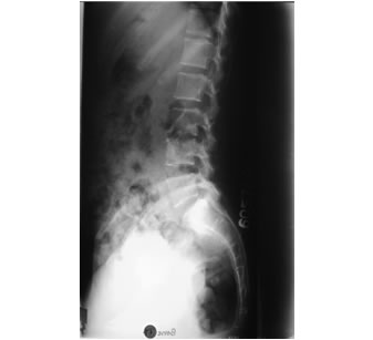
QUESTION:
- Describe the findings in the x-ray.
- What is your diagnosis?
ANSWER:
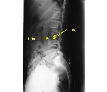
- a) Erosion of the superior end plate of the L4 vertebra and inferior end plate of the L3 vertebra.
b) Narrowed disc space (reduction of the height of the disc space) between L3 and L4 vertebra. No significant vertebral collapse or gibbous. No abnormal soft tissue shadow anterior to the spine. - Tuberculosis of the lumbar spine.
DISCUSSION
Due to the indolent nature of the infection, radiological manifestations are usually advanced when the patient clinically present with symptoms. Erosion of the end plates, of the contiguous vertebra starting from the anterior aspect, is the cardinal radiological feature seen in the plain radiograph. This is due to the spread of the infection from the vertebral body to the disc space. Progressive narrowing of the disc space associated with vertebral collapse leads to the gibbous deformity. (not seen in this X-ray). Para spinal abscess formation is seen as a soft tissue shadow anterior to the spine in the plain radiograph. (not seen in this patient).
Infection may spread, beneath the anterior or posterior longitudinal ligament, or to the spinal epidural space forming epidural abscesses. Multi-detector computered tomography (CT) is helpful in demonstrating the extent of vertebral and disc space destruction and paraspinal abcess formation. Magnetic resonance imaging (MRI) is particularly useful in diagnosing paraspinal and epidural abcesses and sub-ligamentous spread. Differentiation from pyogenic infection is mainly by the erosive changes beginning at the anterior aspect of the vertebra. Predominant involvement of the end plates and the clinical features makes vertebral metastases an unlikely diagnosis.
REFERENCES
- De Backer AI, Mortelé KJ, Vanschoubroeck IJ, et al. Tuberculosis of the spine: CT and MR imaging features. JBR-BTR. 2005;88(2):92–7. [PubMed]
- Turgut M. Spinal tuberculosis (Pott’s disease): its clinical presentation, surgical management, and outcome. A survey study on 694 patients. Neurosurg Rev. 2001; 24(1):8-13. [PubMed]
- Renton P. Periosteal reactions, bone and joint infections, sarcoid. In: David Sutton. Text book of radiology and imaging. 7th ed. Elsevier/Churchill Livingstone; 2002. vol 2 p. 1166-8.
- Wilson DJ, Berendt AR. Bone and soft tissue infections. In: Grainger RG, Allison DJ. Grainger and Allison’s diagnostic radiology. A Text book of medical imaging. 5th ed. Elsevier/Churchill Livingstone; 2008. vol 2 p. 1166.
- Shanley DJ. Tuberculosis of the spine: imaging features. AJR Am J Roentgenol. 1995;164(3): 659-64. [PubMed] [Full text]