
Welcome to the
Malaysian Family Physician, a peer-reviewed open-access journal of family practice
and primary care research.
BRIEF REPORT
NAIL SAMPLING TECHNIQUE AND ITS INTERPRETATION
M Leelavathi1, MN Tzar2
1Department of Family Medicine, Faculty of Medicine, Universiti Kebangsaan Malaysia. (Leelavathi Muthupalaniappen)
2Department of Medical Microbiology and Immunology, Faculty of Medicine, Universiti Kebangsaan Malaysia Medical Centre. (Tzar Mohd Nizam)
Address for correspondence: Assoc Prof Dr Leelavathi Muthupalaniappen, Consultant Family Medicine Specialist, Department of Family Medicine, Faculty of Medicine, Universiti Kebangsaan Malaysia, Jalan Yaacob Latif, Bandar Tun Razak, 56000 Cheras, Kuala Lumpur, Malaysia. Tel: 603 9145 6123, Fax: 603 9173 6680, Email: drleelaraj@yahoo.com
ABSTRACT
The clinical suspicion of onychomyosis based on appearance of the nails, requires culture for confirmation. This is because treatment requires prolonged use of systemic agents which may cause side effects. One of the common problems encountered is improper nail sampling technique which results in loss of essential information. The unfamiliar terminologies used in reporting culture results may intimidate physicians resulting in misinterpretation and hamper treatment decision. This article provides a simple guide on nail sampling technique and the interpretation of culture results.
Leelavathi M, Tzar MN. Nail sampling technique and its interpretation. Malaysian Family Physician. 2011;6(2&3):58-59
INTRODUCTION
Infection of nail apparatus by dermatophytes, non-dermatophyte moulds or yeasts is responsible for 50% of all nail disorders.1 It causes nail deformity such as discolouration, subungual hyperkeratosis and onycholysis. Diagnosis of onychomycosis based on nail morphology alone would help identify about 50% of cases. However, this is not always accurate and nail culture is required for confirmation. Onychomycosis requires prolonged treatment as clinical recovery may take up to one year due to slow growth of nails. Treatment may not necessarily provide clinical response and in addition may cause potential harm. Hence in the absence of nail culture, differentiation between incorrect clinical diagnosis and treatment failure would prove to be difficult.2
Accurate laboratory diagnosis of onychomycosis depends on the quality of sample collection, transportation and reporting by microbiologist. Physicians are often faced with challenges in obtaining adequate, good quality samples and interpretation of culture results as it is frequently dismissed as a trivial procedure.
METHOD OF COLLECTION
Site selection for sampling depends on clinical suspicion of the possible pathogen. In tropical regions where moulds are more likely to cause onychomycosis, nail plate, nail bed and subungual debris provide better specimens for culture. If candida is suspected, the sample is best taken from the proximal and lateral edges of the nail. Aseptic technique prevents overgrowth of contaminants which may suppress growth of pathogenic fungi.3 Adequate sampling increases chances of obtaining positive results compared to smaller amounts as the sampled section of nail may have scanty pathogens. Adequate amount of sample is shown in Figure 1a. A large nail cutter (nail pliers or nipper for thick toenails), scalpel blade for scraping nail plate and a spoon excavator or any small blunt instrument (used in pedicure) can be used for collection of subungual debris. Tools required for sample collection are shown in Figure 1b and a stepwise technique for specimen collection is shown in Table 1. Nail specimens should ideally be cut about 2 to 3 mm thickness.4 Recent studies have shown that the micro-drilling technique for nail sampling provides more accurate diagnosis.5,6 However, the lack of availability of micro-drilling equipment and skills for this technique may be a limiting factor. Hence in most settings, the traditional method of sampling using nail cutter remains the standard practice. Once collected, nail specimen is transported in Petri dishes or small paper envelopes.
Figure 1a: Shows adequate amount of sample
Figure 1b: Shows the tools required for nail sample collection
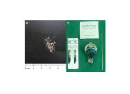 |
Table 1: Steps for specimen collection in onychomycosis
| Steps | Method |
|---|---|
1 |
Stop antifungal agents both systemic and topical, one week before sampling |
2 |
Wipe the toe and nail with moist gauze dipped in saline or distilled water (removes dirt and dust) |
3 |
Clean the discoloured or dystrophic nail plate and nail folds with 70% alcohol (removes coexisting bacteria) |
4 |
Small pieces of nail plate clipping (2 to 3 mm thickness), scrapings of nail bed and sub-ungual debris is collected in a dry paper envelope or agar plate culture medium* |
5 |
Specimen is labelled indicating the site of sampling (finger or toe) |
6 |
Patient information is recorded in the microbiology lab investigation form and transported with the specimen (preferably within 2 hours to ensure optimum culture yield) |
*instruments should be cleaned with alcohol swab and allow to dry before next specimen collection
PROCESSING OF NAIL SAMPLE
Nail specimen should ideally be processed within two hours of collection for optimal results.3 A part of the specimen is subjected to direct microscopy using 20% KOH wet mount and the rest is cultured on Sabouraud dextrose agar. The combination of these two procedures is required to improve accuracy of diagnosis. Sabouraud dextrose agar is pre-mixed with chloramphenicol and cycloheximide to discourage bacterial growth and cultured at 30ºC. Agar plates are reviewed for growth on a daily basis up to four weeks as fungi are slow growing pathogens. Presence of growth on agar plate is subjected to further testing for species identification.
INTERPRETATION OF CULTURE
Culture results are generally reported as positive, mixed growth, contamination or no fungal growth. Positive culture is reported according to the species isolated e.g. non-dermatophyte fungi are reported as aspergillus, fusarium or penicillium sp., yeasts as candida or trichosporum sp. and dermatophyte fungi as Microsporum or trichophyton sp. Mixed growth may represent a true mixture of pathogens or improper collection technique. Contamination suggests improper sample collection or processing. The absence of fungal growth may either represent true absence or a false negative result. About 30-50% of samples may be falsely negative secondary to inadequate sampling or collection of non-viable fungal elements at the distal portion of the nail. Repeat sampling is required if clinical suspicion of onychomycosis is high.7
The isolation of non-dermatophyte fungi may represent laboratory contaminants rather than primary pathogen and hence it poses a dilemma in treatment decision. Non-dermatophyte fungi are non-keratolytic pathogens but are capable of nail invasion resulting in dystrophy and onychomycosis in about 2-12% cases.8,9 In these cases, repeated cultures yielding the same pathogen, heavy growth and positive KOH testing provides a clue to the causative pathogen. However, these findings must be correlated with clinical features.4
CONCLUSION
Adequate nail sample collection using appropriate technique, standardised laboratory processing and culture are essential steps to ensure accurate diagnosis of onychomycosis. It guides clinicians to make decision for treatment initiation or repeat sampling if required. It is beyond doubt that identification of the pathogen by culture and clinical correlation is of utmost importance in guiding physicians to achieve optimal treatment outcome.
AUTHOR’S INFORMATION
The author is a registered Family Medicine Specialist with special interest in dermatology. She trained in dermatology at Kuala Lumpur General Hospital and obtained the Graduate Diploma in Family Practice Dermatology from the National Skin Centre in Singapore.
REFERENCES
- Mügge C, Haustein UF, Nenoff P. Causative agents of onychomycosis - a retrospective study. J Dtsch Dermatol Ges. 2006;4(3):218-28. [PubMed]
- Roberts DT, Taylor WD, Boyle J. Guidelines for treatment of onychomycosis. Br J Dermatol. 2003;148(3):402-10. [PubMed]
- Chaya AK, Pande S. Methods of specimen collection for diagnosis of superficial and subcutaneous fungal infections. Indian J Dermatol Venereol Leprol. 2007;73(3):202-5. [PubMed]
- de Berker D. Clinical practice. Fungal nail disease. N Engl J Med. 2009;360(20):2108-16. [PubMed]
- Shemer A, Trau H, Davidovici B, et al. Collection of fungi samples from nails: comparative study of curettage and drilling techniques. J Eur Acad Dermatol Venereol. 2008;22(2):182-5. [PubMed]
- Mochizuki T, Kawasaki M, Tanabe H, et al. A nail drilling method suitable for the diagnosis of onychomycosis. J Dermatol. 2005;32(2):108-13. [PubMed]
- Ellis DH. Diagnosis of onychomycosis made simple. J Am Acad Dermatol. 1999;40(6 Pt 2):S3-8. [PubMed]
- Moreno G, Arenas R. Other fungi causing onychomycosis. Clin Dermatol. 2010;28(2):160-3. [PubMed]
- Mandell GL, Bennett JE, Dolin R. Principles and practice of infectious diseases. 7th ed. vol 2 chap 267. Philadelphia: Churchill Livingstone Elselvier; 2010. p. 3353.