
Welcome to the
Malaysian Family Physician, a peer-reviewed open-access journal of family practice
and primary care research.
TEST YOUR KNOWLEDGE
Respiratory Clinics
COUGH, HAEMOPTYSIS AND INCREASING BREATLESSNESS IN A 55-YEAR-OLD CACHEXIC MALE
R Khajotia MBBS(Bom), MD(Bom), MD(Vienna), FAMA(Vienna), FAMS(Vienna)
Associate Professor in Internal Medicine and Pulmonology, International Medical University Clinical School, Seremban, Negeri Sembilan, Malaysia. (Rumi Khajotia)
Address of correspondence: Dr Rumi Khajotia, Associate Professor in Internal Medicine and Pulmonology, International Medical University Clinical School, Jalan Rasah, 70300 Seremban, Negeri Sembilan, Malaysia. Tel: 06-7677 798, Fax: 06-7677 709, Email: rumi@imu.edu.my
Khajotia R. Cough, haemoptysis and increasing breathlessness in a 55-year-old cachexic male. Malaysian Family Physician. 2011;6(2&3):87-88
CASE DESCRIPTION
A 55-year-old man came to the outpatient department with complaints of cough, purulent expectoration and breathlessness off and on since the past three years. Since the past two weeks, he has noticed blood in his sputum each time he coughed. He also complains of weight loss of 4 kg accompanied by loss of appetite, over the past three months. He gives history of pulmonary tuberculosis three years ago, for which he took treatment for six months. On examination, the patient appears cachexic. He is tachycardic and tachypnoeic with a pulse rate of 96 beats per minute and a respiratory rate of 32 breaths per minute, respectively. He is dusky in appearance and his blood pressure (BP) is 134/82 mmHg. On examination of his chest, the left hemithorax appears to be “flattened” as compared to the right side. The trachea is shifted to the left side and the left hemithorax also moves significantly less as compared to the right. On percussion, there is a dull note over the entire left side of the chest both anteriorly and posteriorly, while a normal resonant note is heard over the right side. On auscultation, there is diminished air entry over the entire left hemithorax with tubular bronchial breath sounds heard over the left infraclavicular and interscapular regions. Vocal resonance is diminished over the entire left hemithorax except in the left infraclavicular and interscapular regions, where it is increased. Auscultatory findings are normal over the right side. A chest radiograph is done (Figure 1).
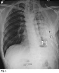
Figure 1: Left fibrothorax with calcification of visceral pleura and trapped lung
- Interpret the chest radiographic findings.
- Based on the history, clinical examination and radiographic findings, what is the likely clinical diagnosis?
- What is the likely aetiological diagnosis of this condition?
- Which further investigations would you perform in this patient?
- A needle-aspiration is suggested on the left side in order to reexpand the partially collapsed lung: Is the procedure justified?
- What is the treatment of choice in this complicated condition?
ANSWER:
- The chest radiograph (Figure 1) shows a significantly contracted left hemithorax with compensatory hyperinflation on the right side. The trachea is shifted to the left side. There is significant pleural thickening on the left side. The left lung appears to be partially collapsed with a thickened visceral pleura surrounding it (black arrows). A few areas of cavitation are seen on the left side (white arrows). The heart is shifted to the left side and the left cardiac contour is not visible.
- “Left-sided fibrothorax” with significant destruction of the left lung parenchyma (as evidenced by multiple cavitations) and partial left lung collapse, suggestive of a “trapped lung” on the left side.
- Pulmonary Tuberculosis with reinfection or reactivation. To look for multi-drug resistance (MDR-TB) or extensive drug resistance (XDR-TB).
- Sputum for AFB smear and culture. (If AFB culture is positive, to undertake drug-sensitivity testing for primary and reserve anti-tuberculosis drugs)
Sputum for TB-PCR (to detect live bacilli quickly, within 48 hours)
Sputum for bacterial culture and sensitivity
Full blood count, ESR, fasting blood sugar levels
Serum gamma-interferron levels (determines the presence of active tuberculous infection)
Electrocardiogram (ECG)
Arterial blood gas estimation
HIV-1 testing
HRCT Thorax
Pulmonary Function Testing (PFT)
Bronchoscopy, if necessary
Thracoscopy, if necessary - The procedure would not be justified as the lung on the left side is “trapped” and would be unable to expand by both needle aspiration and intercostal drainage. This is because there is a strong possibility of surrounding fibrosis and adhesions in the left pleural cavity accompanied by thickened visceral and parietal pleura which would prevent the lung from expanding.
- Surgical management: As the entire left lung appears to be virtually destroyed (multiple cavitations), with surrounding fibrosis and thickened pleural surfaces, the surgical treatment of choice is left-sided pneumonectomy, provided the patient is medically fit to undergo surgery.
- If the TB-PCR results are positive, it is suggestive that the patient has active pulmonary tuberculosis. Since the patient has taken anti-tuberculous treatment in the recent past, it is safer to treat the patient as a potential case of MDR-TB (until the culture and drug-sensitivity testing results are available). For this, the patient can be given a combination of drugs from both, the primary and reserve groups of anti-tuberculous medications. List of reserve anti-tuberculosis medication include: kanamycin, capreomycin, ethionamide, cycloserine, ciprofloxacin, ofloxacin, levofloxacin, amikacin and PAS.
- Antibiotics for superadded bacterial infection.
- Antifungal treatment for superadded fungal infection which commonly occurs in tuberculous lung cavities.
- Treat haemoptysis if and when it occurs. Common causes for this include: active tuberculosis and fungal infection.
Palliative management: Chest physiotherapy, postural drainage, high-protein diet and steam inhalation constitute palliative management in this patient.
REFERENCE TEXT FOR RECOMMENDED READING
- Crofton and Douglas’ Respiratory Diseases. 5th ed. Blackwell Science; 2008.