
Welcome to the
Malaysian Family Physician, a peer-reviewed open-access journal of family practice
and primary care research.
TEST YOUR KNOWLEDGE
A MAN WITH MULTIPLE ABDOMINAL CALCIFICATIONS
M Rohsila1, D Nani2, Y Siti Suhaila2
1Department of Radiology, School of Medical Sciences, Universiti Sains Malaysia, Kubang Kerian, Kelantan. (Rohsila Muhamad)
2Department of Family Medicine, School of Medical Sciences, Universiti Sains Malaysia, Kubang Kerian, Kelantan.
(Nani Draman, Siti Suhaila Yusoff)
Address for correspondence: Dr Rohsila Muhamad, Department of Radiology, School of Medical Sciences, Universiti Sains Malaysia, Kubang Kerian, Kelantan, Malaysia. Tel: 609-767 3000/6619, Fax: 609-767 6611, Email: mrohsila@yahoo.com
Rohsila M, Nani D, Siti Suhaila Y. A man with multiple abdominal calcifications. Malaysian Family Physician. 2012;7(1):45-46
CASE HISTORY
A 65-year-old man presented with a dull, aching pain in the left lumbar region for three days duration. The pain was aggravated by movement and associated with low grade fever and vomiting. He had a few episodes of passing out sandy particles in his urine, several days prior to admission. He denied any lower urinary tract symptoms or alteration in his bowel habits.
Physical examination revealed stable haemodynamic status. His blood pressure was 130/70 mmHg with a pulse rate of 96 beats/min. His body temperature was 37.6ºC. He had mild tenderness in the left lumbar region on deep palpation of his abdomen. Kidneys were not ballotable and renal punch was negative. His kidney ureter bladder (KUB) radiograph is shown in the following image (Figure 1).
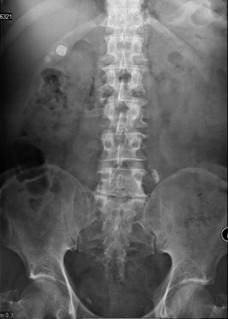
Figure 1
QUESTION:
- Describe the main findings in the radiograph and give reasons for the diagnoses.
- What is the next best radiological investigation to be performed?
ANSWER:
- This is a kidney ureter bladder (KUB) radiograph taken in a supine position. There are multiple well defined opacities at different locations with various sizes and shapes. The right hypochondrium opacities are rounded with a lamella appearance. They are located between the shadow of the liver margin and the right renal outline. The left iliac fossa opacity is tubular in shape and overlies the left transverse process of the fifth lumbar vertebra. Bilateral opacities in the pelvis are located below the level of the ischial spine. Both renal and psoas muscle outlines are seen. Visualised bones are unremarkable. These findings are in keeping with cholelithiasis, left distal ureteric calculus and pelvic phleboliths.
- Ultrasound is the best primary modality to assess gallstones and its complications e.g. choledocholithiasis, cholecystitis or gallstone ileus.1 Multi slice computed tomography (CT) may provide the exact number, size, and location of ectopic stones. It can diagnose gallstone ileus, intestinal obstruction or may reveal direct visualisation of a biliary enteric fistula for therapeutic management of patients.2
Generally, site of the calculus and typical features of each calcification suggest the most likely diagnosis. Cholelithiasis or gallstones are mainly cholesterol stones. Approximately 15-20% of gallstones contain enough calcium to be visible on plain radiographs.1 Radiographically, gallstones are usually rounded in shape with rim/egg shell calcification. Gallbladder stones are usually multiple and sited in the hypochondrium area.
A KUB can often visualise calcium-containing stones in the kidney or ureter, including struvite stones. Uric acid or other purine stones may be radiolucent, and cystine stones often visualize poorly.3 Ureteric stones are usually tubular in shape, conforming to the ureteric lumen. 60% of calcifications along the expected course of the ureter (which overlies the tip of the transverse process of the lumbar vertebra) on the symptomatic side are ureteric stones.
Phleboliths or a calcified vessel wall is common in the lateral part of the pelvis below the level of the ischial spine. Typically, a phlebolith has a radiolucent centre on plain radiograph.4 It is unlikely representing a vesicoureteric junction (VUJ) stone because the VUJ is located above the level of the ischial spine anatomically.
Ureteric calculus is best studied by intravenous urography (IVU) or CT urography. IVU will provide information on renal function, confirm the location of the ureteric stone and assess obstructive hydronephrosis. In addition, CT urography confirms the location of the calculus, detecting other non-visible calculi on plain radiograph, as well as assessing surrounding tissue and kidney morphology. CT will also confirm the presence of the phleboliths in the pelvis.
REFERENCES
- Bortoff GA, Chen MY, Ott DJ, et al. Gallbladder stones: imaging and intervention. Radiographics. 2000;20(3):751-66. [PubMed]
- Lassandro F, Romano S, Ragozzino A, et al. Role of helical CT in diagnosis of gallstone ileus and related conditions. AJR Am J Roentgenol. 2005;185(5):1159-65. [PubMed] [Full text]
- Worcester EM, Coe FL. Nephrolithiasis. Prim Care. 2008;35(2):369-91, vii. [PubMed] [Full text]
- Traubici J, Neitlich JD, Smith RC. Distinguising pelvic phleboliths from distal ureteral stones on routine unenhanced helical CT: is there a radiolucent center? AJR Am J Roentgenol. 1999;172(1):13-7. [PubMed] [Full text]