

Welcome to the
Malaysian Family Physician, a peer-reviewed open-access journal of family practice
and primary care research.
TEST YOUR KNOWLEDGE

Respiratory Clinics
HAEMOPTYSIS, BREATHLESSNESS AND CHEST PAIN IN A 46-YEAR-OLD MAN
R Khajotia, MBBS(Bom), MD(Bom), MD(Vienna), FAMA(Vienna), FAMS(Vienna)
Associate Professor in Internal Medicine and Pulmonology, International Medical University Clinical School, Seremban, Negeri Sembilan, Malaysia. Email: rumi@imu.edu.my
Address of correspondence: Dr Rumi Khajotia, Associate Professor in Internal Medicine and Pulmonology, International Medical University Clinical School, Jalan Rasah, 70300 Seremban, Negeri Sembilan, Malaysia. Tel: 06-767 7798, Fax: 06-767 7709, Email: rumi@imu.edu.my
Khajotia R. Haemoptysis, breathlessness and chest pain in a 46-year-old man. Malaysian Family Physician. 2010;5(2):105-6
CASE REPORT
A 46-year-old man came to the outpatient department with complaints of progressively increasing right-sided chest pain accompanied by tenderness since six months, which even disturbed his sleep. Since the past three months he also noticed breathlessness which has progressively increased and since one week he is breathless even at rest. A week ago, he noticed bright red blood in his sputum. Since then, he has been coughing out fresh blood, about a quarter-cupful a day. On questioning, the patient said that he was a heavy smoker who smoked 30 cigarettes a day for the past 27 years. He worked as a clerk in an office. There was no history suggestive of contact with a tuberculous patient. His family members too were healthy and well. He had no past history of diabetes mellitus or hypertension. On examination, the patient appeared breathless with a respiratory rate of 28 breaths per minute. His pulse rate was 108 beats per minute and the blood pressure was 102/68 mmHg. The trachea was shifted to the left side. There was gross tenderness over the right side of his chest, both anteriorly and posteriorly. Chest wall movements were diminished on the right side and there was a dull note on percussion over the entire right hemithorax, both anteriorly and posteriorly. On auscultation, air entry was absent over the right mid-zone and lower-zone and tubular bronchial breath sounds were heard in the right interscapular region. A chest radiograph was done (Figure 1).
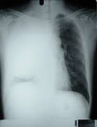
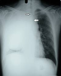
Figure 1 Figure 2
QUESTION:
- Interpret the chest radiograph in Figure 1.
- What do the open arrow and white arrow in the chest radiograph in Figure 2 indicate?
- What is the predominant cause of breathlessness in this patient?
- Based on the history, physical examination findings and chest radiograph, what is the most likely diagnosis in this patient?
- What is the cause of his persistent chest pain?
- What further investigations would you perform to confirm the diagnosis?
- What does the presence of haemoptysis signify in this patient?
- What is the prognosis for this patient?
ANSWER:
- The chest radiograph in Figure 1 shows a large homogenous opacity in the right hemithorax. This opacity has a well-defined lower margin and extends to the periphery, upto the chest wall. The trachea appears to be shifted to the left side and there is compensatory hyperinflation of the left lung.
- The open arrow in Figure 2 shows a sharp “cut-off” of the right main-stem bronchus suggestive of a complete endobronchial obstruction. There is also significant narrowing of the left main-stem bronchus which exhibits a typical “rat-tail” appearance as shown by the white arrow. This is suggestive of an external compression.
- Complete occlusion of the right main-stem bronchus and significant narrowing of the left main-stem bronchus accompanied by significant collapse of the right lung are prime causes of breathlessness in this patient.
- Based on the clinical examination and chest radiographic findings the most likely diagnosis is ‘bronchogenic carcinoma’ involving the right lung.
- The tumour mass appears to have spread to the periphery of the right lung and has significantly infiltrated the right chest wall which is responsible for the persistent, unrelenting chest pain in this patient.
- Other investigations include:
- Sputum examination for malignant cells1
- High resolution CT scan of the chest2
- PET scanning using fluoro-18-2-deoxyglucose (FDG)3
- Bronchoscopic examination for diagnostic and therapeutic purposes4
- Percutaneous tumour biopsy or fine needle aspiration cytology
- Tumour markers
- CT scan of the abdomen and brain to look for secondaries (metastasis) in other parts of the body
- Bone scan
- Colonoscopy, if indicated
- Malignant tumours can be highly vascular. Hence, the presence of frank haemoptysis indicates that the tumour mass has infiltrated into the tracheobronchial tree with an endobronchial extension of the tumour process. Haemoptysis has often been described as the one symptom often prompting more rapid presentation and earlier diagnosis.5
- As the tumour has already infiltrated the chest wall on the right side and also appears to have infiltrated to within 2 cm of the carina with a sharp cut-off of the right main-stem bronchus and significant narrowing of the left main-stem bronchus, the malignancy is in stage IV. Hence the overall prognosis is poor, with a five year survival rate of less than 2%.6
REFERENCES
- Erkilic S, Ozsarac C, Kullu S. Sputum cytology for the diagnosis of lung cancer. Comparison of smear and modified cell block methods. Acta Cytol. 2003;47(6):1023-7. [PubMed]
- Forster BB, Muller NL, Miller RR, et al. Neuroendocrine carcinomas of the lung: clinical, radiologic, and pathologic correlation. Radiology. 1989;170(2):441-5. [PubMed]
- Gould MK, Maclean CC, Kuschner WG, et al. Accuracy of positron emission tomography for diagnosis of pulmonary nodules and mass lesions: a meta-analysis. JAMA. 2001;285(7):914-24. [PubMed] [Full text]
- Arroliga AC, Matthay RA. The role of bronchoscopy in lung cancer. Clin Chest Med. 1993;14(1):87-98. [PubMed]
- Corner J, Hopkinson J, Fitzsimmons D, et al. Is late diagnosis of lung cancer inevitable? Interview study of patients' recollections of symptoms before diagnosis. Thorax. 2005;60(4):314-9. [PubMed] [Full text]
- Ries L, Eisner M, Kosary C. Cancer statistics review, 1975–2002. National Cancer Institute; 2005. [Link]