

Welcome to the
Malaysian Family Physician, a peer-reviewed open-access journal of family practice
and primary care research.
TEST YOUR KNOWLEDGE

Respiratory Clinic
DRY COUGH AND SEVERE WEIGHT LOSS
R Khajotia MBBS(Bom), MD(Bom), MD(Vienna), FAMA(Vienna), FAMS(Vienna)
Associate Professor in Internal Medicine and Pulmonology, International Medical University Clinical School, Seremban, Negeri Sembilan, Malaysia. (Rumi Khajotia)
Address of correspondence: Dr Rumi Khajotia, Associate Professor in Internal Medicine and Pulmonology, International Medical University Clinical School, Jalan Rasah, 70300 Seremban, Negeri Sembilan, Malaysia. Tel: 06-767 7798,
Fax: 06-767 7709, Email: rumi@imu.edu.my
CASE REPORT
A 55-year-old male came to his family physician complaining of cough since six months. The cough was non-productive. It was persistent throughout the day and also at night, which disturbed his sleep. Over the past three months, he also noticed he had lost 20 kg weight. There was no history of tuberculosis in the past or amongst his family members or friends. There was no history of chest pain, fever, breathlessness or haemoptysis. He was neither a diabetic nor a hypertensive. He was a chronic smoker, smoking 30 cigarettes a day for the past 25 years. He did clerical duties in an office. On examination, the patient appeared cachexic. His vital parameters were normal. His pulse rate was 76 beats per minute, respiratory rate was 22 breaths per minute and his blood pressure was 128/84 mmHg. He was not cyanosed and his accessory muscles of respiration were not active. On examination of his chest, it was observed that both sides of his chest moved equally well with respiration. A dull note on percussion was heard in the right interscapular region. Rest of the lung fields were normal on percussion. On auscultation, localised rhonchi and tubular bronchial breath sounds were heard in the right interscapular region, posteriorly. Normal vesicular breath sounds were heard over rest of the lungs fields. The patient was immediately referred to a specialist. A chest radiograph was urgently done (Figure 1) and the patient was admitted to hospital for further investigations and management.
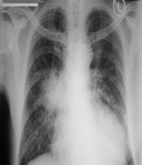
Figure 1: Chest radiograph taken at time of admission
QUESTION:
- What do you see on the chest radiograph?
- What are the likely differential diagnoses based on this chest radiograph?
- Based on the patient’s history and chest radiographic findings, what is the most likely final diagnosis in this patient?
- In order to confirm the diagnosis, what other investigations would you perform in this patient?
- What are the treatment options available for this patient?
ANSWER:
- The chest radiograph shows a diffuse opacity 5 x 4 cm in size in the right parahilar region. The opacity has ill-defined outer borders (Figure 2, black arrows) which appear to infiltrate into the surrounding pulmonary parenchyma. The left hilar region appears slightly prominent.
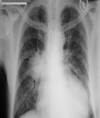
Figure 2: A diffuse opacity 5 x 4 cm in size is seen in the right parahilar region. The opacity has ill-defined outer borders (black arrows) which appear to infiltrate into the surrounding pulmonary parenchyma. The left hilar region appears slightly prominent.
- The likely differential diagnoses include:
- Consolidation in the right parahilar region due to bacterial, tuberculous (primary), viral or fungal (histoplasmosis, blastomycosis) infection
- Right hilar lymphadenopathy due to infection or malignancy (hodgkin’s/non-hodgkin’s lymphomas)
- Right hilar tumour mass due to a malignant process (bronchogenic carcinoma)
- Metastatic lymph node enlargement
- Hilar lymphadenopathy due to chronic use of drugs such as phenytoin or dilantin
- Hilar lung abscess
- Tumours such as lipoma, fibroma, liposarcoma, fibrosarcoma and mediatinal teratoma
- Eosinophilic granuloma
- This patient has had a persistent dry cough over six months followed by severe weight loss of 20 kg over a rather short duration of time (three months). There is no history suggestive of infection such as fever, purulent expectoration or pleuritic chest pain. There is no history of tuberculosis or contact with a tuberculous patient. He is a chronic heavy smoker. There is no history of chronic use of drugs such as phenytoin or dilantin. On examination, a dull note on percussion, localised rhonchi and tubular bronchial breath sounds are observed in the right interscapular region, posteriorly. The chest radiograph shows a large rounded opacity in the right parahilar region which has ill-defined borders which appear to infiltrate into the surrounding pulmonary parenchyma. There is no air-fluid level seen within the opacity. There is no evidence of interstitial or pleural involvement. The left hilum too appears to be slightly prominent. In view of these findings the most likely diagnosis is a right hilar tumour mass due to Primary Bronchogenic Carcinoma (Lung Cancer).
- Other essential investigations include:
- Sputum for malignant cells (4 days)1
- High-resolution CT-scan of the chest
- Pulmonary Function Testing
- Fibre-optic/Video-bronchoscopy2
- Tumour markers
- CT-scan of the abdomen and brain to look for metastasis
- If the tumour is periferal (posteriorly located), to consider percutaneous transthoracic fine-needle biopsy of the lesion
- Other supportive investigations include:
Total white blood count to rule out superadded infection
Stool examination to look for malaena (colonic malignancy)
-
- Surgery.3 Radical tumour resection is possible if the tumour mass is central with little or no mediastinal involvement. Contraindications for radical resection include histological type (SCLC), diffuse mediastinal involvement, tumour infiltration within 2 cm of the carina, chest wall infiltration, phrenic nerve involvement, malignant pleural effusion, distant metastasis, poor general condition of the patient, poor lung functions and co-morbid conditions which may contraindicate surgery.
- Palliative chemotherapy and radiotherapy3 are usually reserved for cases where surgery is palliative or contraindicated. They help to reduce the tumour burden and thereby improve quality of life.
- Pain management is essential to improve the quality of life in these patients especially in cases where there is chest wall infiltration by the tumour and mediastinal involvement.
- Supportive care is essential. It includes a balanced diet and regular feeds.
- In terminal cases, management of acute breathlessness may be necessary. This may involve the use of endotracheal intubation, invasive ventilation and tracheo-bronchial stenting.
REFERENCES
- 1. Erkilic S, Ozsarac C, Kullu S. Sputum cytology for the diagnosis of lung cancer. Comparison of smear and modified cell block methods. Acta Cytol. 2003;47(6):1023-7. [PubMed]
- Arroliga AC, Matthay RA. The role of bronchoscopy in lung cancer. Clin Chest Med. 1993;14(1):87-98. [PubMed] Reference text for recommended reading:
- European Society for Medical Oncology. Textbook of Lung Cancer. 2nd ed. Edited by Heine Hansen. United Kingdom:Informa Healthcare; 2008.